Gallery
Here we present a selection of projects where CIPA experts have been involved in image processing and analysis. Click on images to view them in full resolution.
Images marked "CC-BY-4.0" are distributed under the terms of the Creative Commons CC-BY-4.0 license, which permits unrestricted use, distribution, and reproduction in any medium, provided the original work is properly cited.
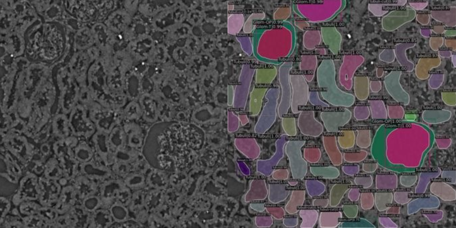
About: A machine learning based segmentation and quantification study to estimate the size of Bowman’s space. Bowman’s space is the difference between outer perimeter and Tuft in a glomeruli. Such approximations can reveal tendencies toward having or developing cancer.
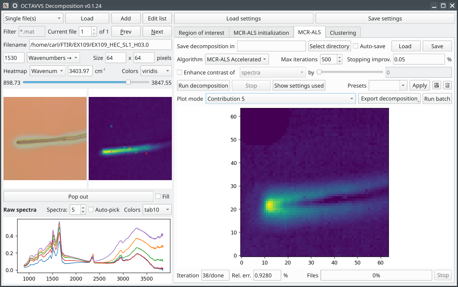
Publication: OCTAVVS: A graphical toolbox for high-throughput preprocessing and analysis of vibrational spectroscopy imaging data. C Troein, S Siregar, M Op De Beeck, C Peterson, A Tunlid and P Persson. Methods and Protocols, 2020. doi.org/10.3390/mps3020034
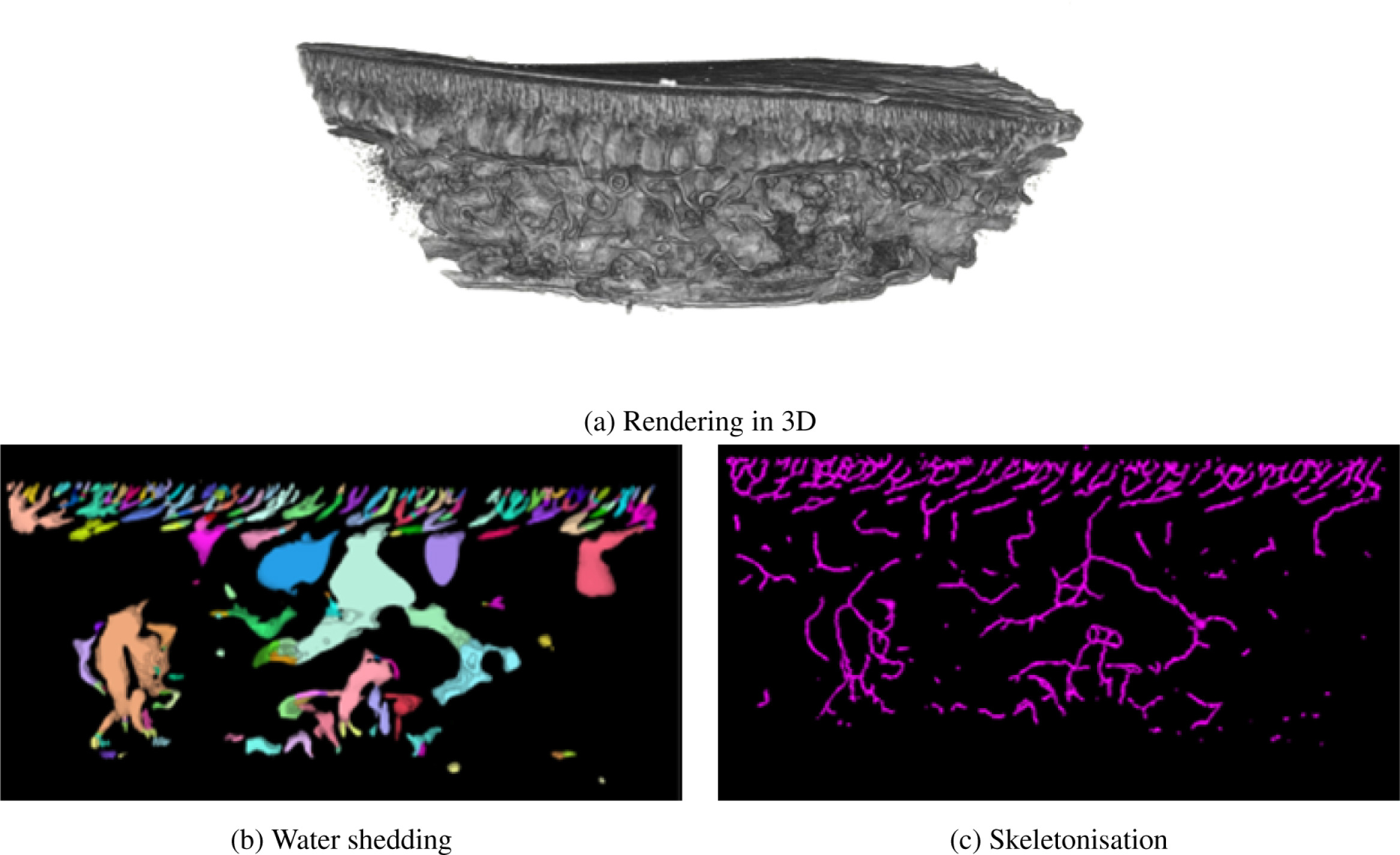
Publication: Towards multiscale X-ray tomographic imaging in membrane science—A perspective. G Rudolph-Schöpping, E Larsson, TN Pingel, M Guizar-Sicairos, P Villanueva-Perez, S Hall and F Lipnizki. Journal of Membrane Science, 2023. doi.org/10.1016/j.memsci.2023.122245
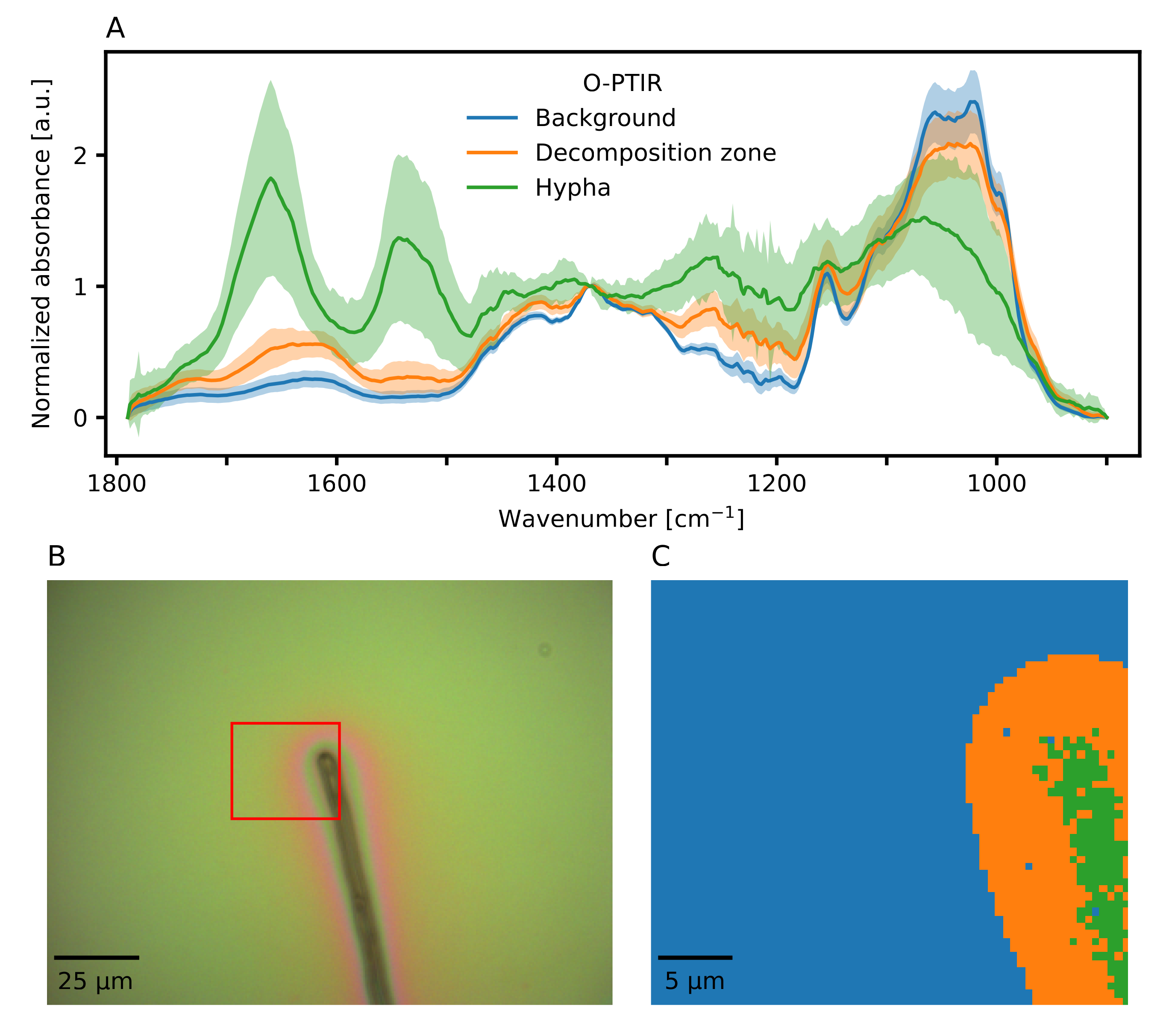
Publication: Elucidating fungal decomposition of organic matter at sub-micrometer spatial scales using optical photothermal infrared (O-PTIR) microspectroscopy. M Op De Beeck, C Troein, C Peterson, A Tunlid and P Persson. Applied and Environmental Microbiology, 2024. doi.org/10.1128/aem.01489-23
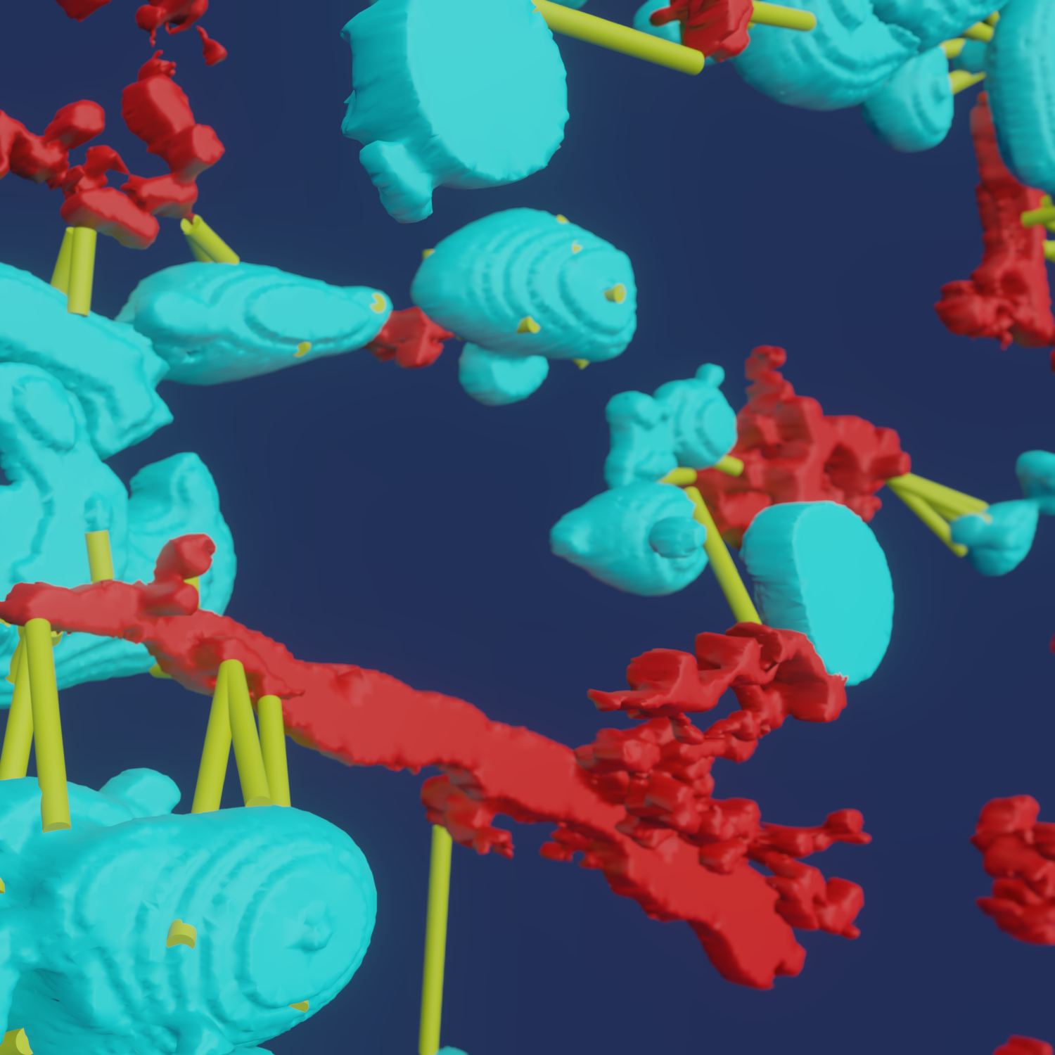
About: CIPA provides expertise in 3D data processing, modeling, raytracing etc.
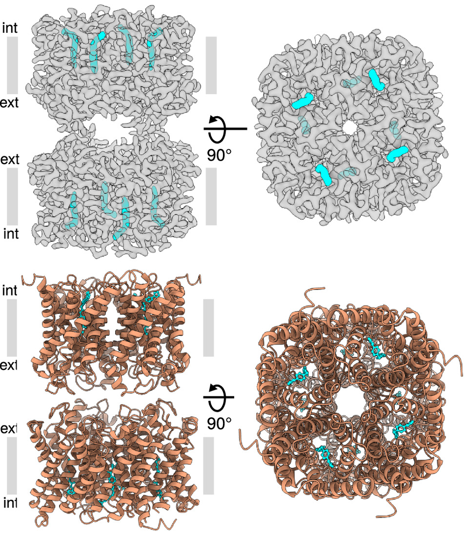
Publication: Molecular basis for human aquaporin inhibition. P Huang, H Åbacka, CJ Wilson, ML Wind, M Rützler, A Hagström-Andersson, P Gourdon, BL de Groot, R Venskutonytė and K Lindkvist-Petersson. PNAS, 2024. doi.org/10.1073/pnas.2319682121